24 October 2024
9:00—17:30
Great Lecture Hall / Velká posluchárna (2. LF UK)
After the capacity is full, it is still possible to watch the programme without interactive aids.
Second Faculty of Medicine, Charles University in collaboration with Institute of Cardiovascular Sciences, University College London (UCL) and Great Ormond Street Hospital (GOSH), London
Presented by: Prof. Andrew Cook, Dr Claudio Capelli, Endrit Pajaziti, Prof. Jan Marek, Prof. Silvia Schievano and Dr Beatrice Bonello, University College London (UCL) and Great Ormond Street Hospital London (GOSH), UK.
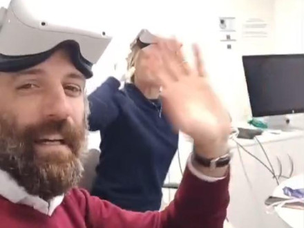
This innovative one-day course provides an in-depth review of normally and abnormally structured heart combining Anatomy Labs and Echocardiographic imaging using novel Virtual Reality (VR) educational program.
Led by a multi-disciplinary team of consultant morphologists and engineers from GOSH and UCL, this unique training offers an in-depth review of structurally normal heart and examples of congenital heart abnormalities in order to address the challenges of lesions with marked anatomical variability. 2D echocardiographic imaging (first-line imaging in cardiology) combined with 3D anatomical images provide a practical approach to how 2D imaging interprets 3D cardiac structures. This course also offers the possibility to familiarize with computer modelling and simulations for a multidisciplinary enrichment of understanding of large variety of congenital heart abnormalities.
Learning objectives:
- To understand normal cardiac anatomy
- To understand the anatomical variability in congenital heart abnormalities
- To understand cardiac anatomy using Echocardiography (first choice imaging modality in Cardiology)
- To familiarise with VR technology for future learning and planning
- To familiarise with computer modelling and simulations
Who should attend?
The course is aimed at advanced Medical students (4th–6th year), Junior doctors and Cardiology trainees specialising in Paediatric Cardiology and Adult Cardiology in Congenital Heart Diseases. No previous VR experience is required.
Register here.
Course Program:
| 9:00 | Arrival & Registration | |||||||||||||||||||||||||
| 9:15–9:30 |
Welcome and introduction Prof. Jan Marek and Dean Prof. Marek Babjuk |
|||||||||||||||||||||||||
| 9:30–10:15 |
Anatomy basics of ASD, VSD and DORV Prof. Andrew Cook |
|||||||||||||||||||||||||
| 10:15–11:00 |
Principles of Echo assessment in ASD, VSD and DORV Dr Beatrice Bonello |
|||||||||||||||||||||||||
| 11:00–11:20 | —coffee break— | |||||||||||||||||||||||||
| 11:20–12:10 | Hands-on VR experience* #1: Normal heart | |||||||||||||||||||||||||
| 12:10–13:00 | Hands-on VR experience* #2: ASD | |||||||||||||||||||||||||
| 13:00–14:00 | —lunch— | |||||||||||||||||||||||||
| 14:00–14.20 |
Introduction to VR in cardiology Dr Claudio Capelli |
|||||||||||||||||||||||||
| 14:20–14:45 |
Modelling in CHDs – Challenges and future opportunities Prof. Silvia Schievano |
|||||||||||||||||||||||||
| 14:45–15:30 | Hands-on VR experience* #3: VSD | |||||||||||||||||||||||||
| 15:30–15:45 | —Coffee Break— | |||||||||||||||||||||||||
| 15:45–16:30 | Hands-on VR experience* #4: DORV | |||||||||||||||||||||||||
| 16:30–17:30 | Networking | |||||||||||||||||||||||||
|
* Each hands-on VR experience is able to host 20 participants. With max 80 participants we suggest the following grouping |
||||||||||||||||||||||||||
During the whole day, VR stations will be available for 1-to-1 testing |
||||||||||||||||||||||||||
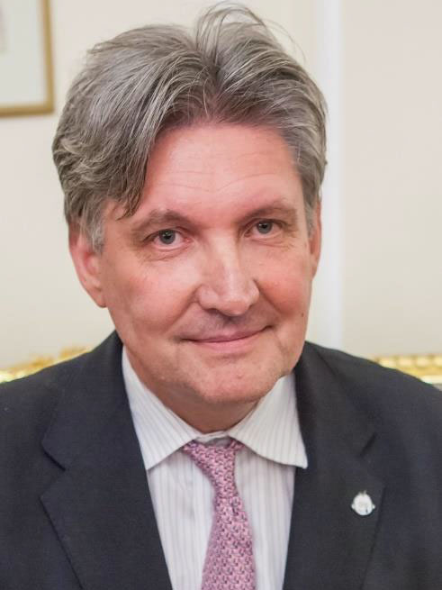 Professor Jan Marek is a Consultant Cardiologist with expertise in paediatric and prenatal cardiology and cardiovascular imaging. He has been working at Great Ormond Street Hospital for Children (GOSH) since 2005 after a previous 20-year professional career at Motol University Hospital, Prague, Czech Republic. Jan is a Professor at the Institute of Cardiovascular Sciences at University College London (UCL) and Charles University, Prague.
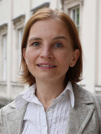 Silvia Schievano is a Professor of Biomedical Engineering at University College London and Great Ormond Street Hospital for Children. Over the past 20 years, she has established and integrated a team of engineers and computer scientists within the hospital, focused on improving treatments for congenital diseases. Her research centres on congenital heart defects and craniofacial abnormalities, employing engineering methodologies to investigate these conditions and driving the development of new medical devices and procedures.
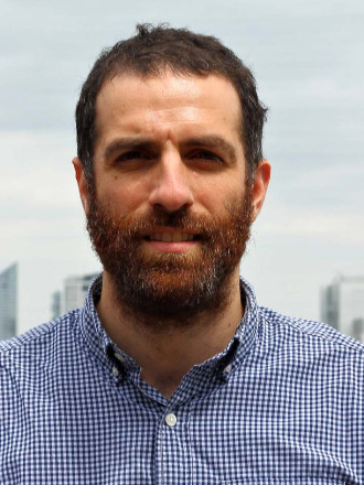 Dr Claudio Capelli is a Senior Researcher in biomedical engineering at University College London (UCL) and Great Ormond Street Hospital (GOSH). After more than 15 years in this field, Claudio remains passionate about cardiac modelling and simulations particularly in supporting clinical decision-making process. He shares this interest with many academic and clinical collaborators all around the world.
 Prof. Andrew Cook leads the Centre for Morphology & Structural Heart Disease at UCL’s Institute of Cardiovascular Science / Great Ormond Street Hospital, now based at the GOSH/UCL Zayed Centre for Research into Rare Disease in Children, London, UK.
He is active in research, education, training with expertise in the structural architecture of the heart during fetal development, in congenital & acquired adult disease, and is the founder of The Heart Academy.
Current research focusses on Organ-to-Cell Molecular Imaging (OCMI) using Hierarchical Phase Contrast Tomography (HiP-CT); Deep-phenotyping of congenital and structural heart disease using synchrotron imaging and HiP-CT; the use of VR and 3D immersive environments for dissemination & education; and structural anatomy for device design.
Související
Zobrazovací metody se rozvíjejí nejrychleji ze všech specializací v medicíně
S prof. Janem Markem o unikátním kardiologickém výukovém programu a nejnovější technice zobrazení (nejen) srdce
Synchrotron X-rays Are A Billion Times More Powerful than Standard X-rays
Prof. Andrew Cook, a heart anatomist and guest lecturer at the workshop on cardiac imaging, has been involved in the development of the world´s latest hearth-imaging method. An interview.

