Info

Fotografie: Hynek Glos (UK)

Epilepsy Surgery: The Greatest Joy Is a Child Without Seizures

Epilepsy Surgery: The Greatest Joy Is a Child Without Seizures

Epilepsy is a very burdensome disease that reduces the quality of life and can sometimes be life-threatening. Epilepsy surgery can eliminate seizures in patients for whom pharmacological treatment is ineffective. In 2024, the Department of Paediatric Neurology at the Second Faculty of Medicine, Charles University, and Motol University Hospital operated on their 500th pediatric patient – eighty percent of them stopped having seizures. The successful program is supported by teams of neurologists, bioengineers, neurosurgeons, psychologists, and radiologists, among others.
Martin Kudr, MD, PhD

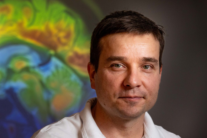
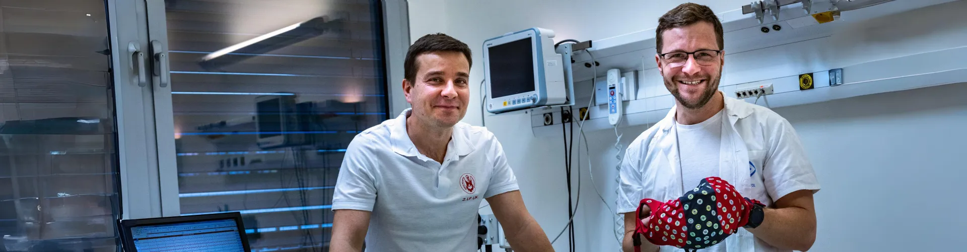
is a physician-epileptologist and head of the paediatric epilepsy surgery programme at the Department of Paediatric Neurology at the Second Faculty of Medicine, Charles University, and Motol University Hospital. His main research interests include clinical science in the field of epileptology and epilepsy surgery, especially functional imaging, SEEG, and insular epilepsy.

Matyáš Ebel, MD, BSc

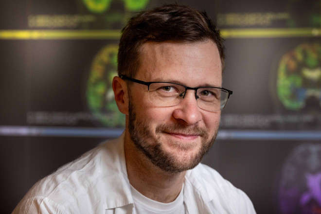
is a secondary physician and doctoral student at the Department of Paediatric Neurology at the Second Faculty of Medicine, Charles University, and Motol University Hospital. He studied General Medicine at the Second Faculty of Medicine, Charles University, and Cybernetics and Robotics at the Czech Technical University. He specialises in epilepsy surgery with an emphasis on the analysis of intracranial EEG (iEEG), high-density EEG (HD-EEG), and neuroimaging, especially in patients with tuberous sclerosis.

The paediatric epilepsy surgery programme has been led by Prof. Pavel Kršek at the Department of Paediatric Neurology at the Second Faculty of Medicine, Charles University, and Motol University Hospital for 25 years. It provides care for children with epilepsy from the Czech Republic, Slovakia, and other European countries. In 2024, the 500th patient was operated on, and the success rate of eliminating seizures increased to 80 percent. The Epileptology Research Centre (EpiReC) is involved in the implementation of innovative diagnostic and therapeutic procedures.

How do epilepsy foci form in the brain?

Martin Kudr: Most often due to congenital malformations of cortical development, benign tumours, less commonly due to brain damage around the time of birth, or, for example, in tuberous sclerosis. The epileptogenic zone, where brain cells are activated more than they should be, is usually one. However, in some severe patients, typically with focal cortical dysplasia type 1, it can be scattered over a larger part of the brain.

Matyáš Ebel: Patients with tuberous sclerosis can also have multiple seizure-generating foci. However, usually, only one place, the hub of the network, is the most active, and that is what we try to locate and surgically disrupt.

If you remove the affected cells, what happens to the resulting space? One of your patients had about a quarter of her brain removed this way.

M. K.: It fills with cerebrospinal fluid, which normally cushions impacts throughout the brain, and scarring forms at the edge of the resection. This does not limit the patient in the long term; within two months after surgery, they can play football, for example. The scar is just a cosmetic defect; there may be an unevenness or a groove in the bone underneath. Paradoxically, cognitive performance usually improves after removing part of the brain because seizures no longer harm the brain.

Do epileptogenic zones correspond to functional brain centres? For example, does epilepsy of the motor centre manifest in the limbs?

M. E.: It can be. The areas of the brain where seizures begin can be close to so-called eloquent areas, which are responsible for higher functions – movement, speech, attention, or memory – or sometimes they can even overlap. If a patient has cortical dysplasia near the motor centre for a limb, seizures can manifest as jerking of that limb. When planning surgery, it is crucial to determine exactly where the motor pathways run, prepare imaging data for the surgeon, and then proceed as gently as possible, with motor monitoring.

Why is it so important to respond and treat early, especially in the youngest infants?

M. K.: The developing brain is more susceptible to damage from severe epilepsy. If we respond early, we give the brain a chance to minimise damage, and the child can develop as normally as possible. In epileptology, the paradigm is currently changing worldwide. We do not wait to see if epilepsy progresses to a very severe form – if we see an opportunity, we operate on the child earlier, often already in infancy.

What examinations must a patient undergo to learn about all the significant zones and pathways?

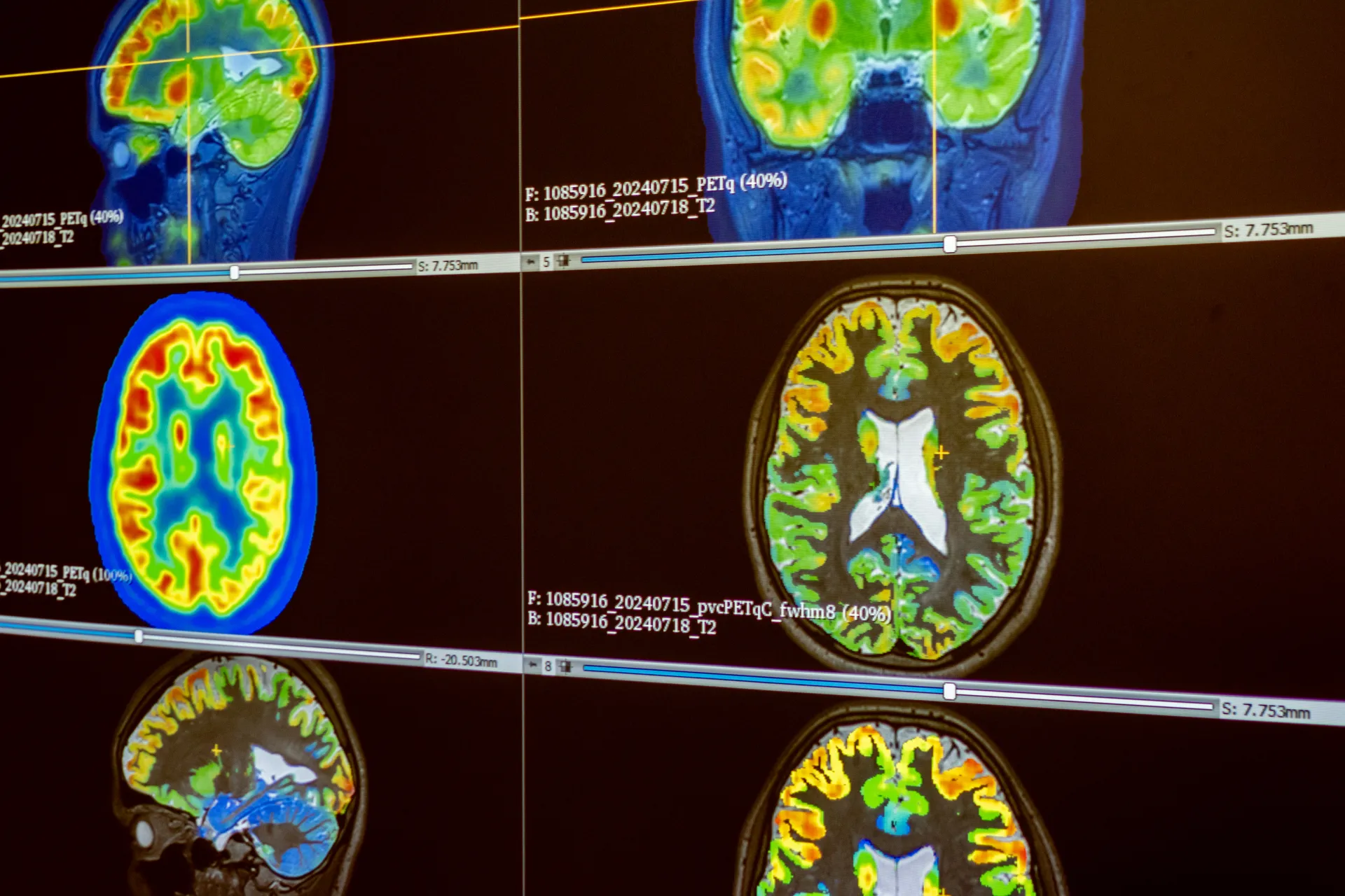
M. E.: The first in the basic range of examinations is video-EEG. This determines brain activity outside of seizures and ideally during a seizure, while we also record the patient on video and evaluate the course of the seizure. Next is magnetic resonance imaging, FDG-PET modified by the pvc-PET method, and neuropsychological examination. If necessary, we supplement with other functional neuroimaging or electrophysiological examinations.

Let's stop at pvc-PET, because engineers from the Czech Technical University played a significant role in its development. The team led by Radek Janča improved the older FDG-PET imaging to better map the metabolic activity of tumour cells. How does it work?

M. K.: Pvc-PET significantly improves FDG-PET itself. The principle is that in the brain area where less sugar is consumed, there is usually also an epileptogenic zone. PET shows this, but the images are coarse and difficult to navigate. One improvement is that PET images are fused with magnetic resonance imaging, which significantly refines the localisation of the epileptogenic zone.

M. E.: To this the limiting condition is added is that the brain's metabolism is mainly generated by the cerebral cortex. This removes the PET signal from the white matter, further refining the examination. Such images are also easier to view.

The pvc-PET imaging method relies on the intersection of information from multiple methods. Archive of the Department of Paediatric Neurology.
Is it used only at your facility in Motol?

M. K.: Pvc-PET was not invented in the Czech Republic, but we are the only epilepsy centre that uses it routinely in ninety-five percent of patients. Studies are currently underway in which we want to demonstrate its advantages.

Can PET, based on some machine learning, determine epileptogenic zones on its own?

M. K.: A human will always be responsible for interpreting the images. However, the key point is that we do not have a single golden method for localising the epileptogenic zone. We rely on the intersection of information from multiple methods. I am not aware that artificial intelligence currently intervenes in the overall evaluation process, although it is starting to help with magnetic resonance imaging.

M. E.: The advantage of machine learning is evident with a larger amount of data than a normal mortal can process. For example, with patients with implanted stereo-EEG electrodes, you evaluate up to one hundred forty contacts. Moreover, they are placed individually in different parts of the brain, and you evaluate almost in real-time because hospitalisation ends with resection. Machine learning helps us detect abnormalities where they cannot be captured by the eye.

Can it be said that the machine surpasses human cognitive ability here?

M. E.: Essentially, yes. But it is just another modality that we must verify again. We certainly cannot rely on the machine yet. We are glad that we understand what the data mean, based on which we make decisions.

You mentioned stereo electroencephalography, SEEG, in which electrodes are implanted in the brain. What is it advantageous for and for which patients?

M. K.: Stereo-EEG is a type of so-called invasive monitoring. It originated in the 1960s in France and has dominated epilepsy surgery worldwide since the late 2000s. I consider it the ultimate method that can best determine where seizures originate and can also help localise eloquent areas.

Thin electrodes, eight-tenths of a millimetre wide, with contacts placed every two millimetres, are inserted into the brain. This allows us to record EEG signals directly from the tissue, which is very precise.

We use the SEEG method in about a quarter of our operated patients. It is overall more demanding for the patient and the entire team, but it is not dangerous.

What does the whole process look like?

M. K.: After quite time-consuming planning of electrode paths, neurosurgeons insert the electrodes into the brain in precisely defined trajectories. The next day, we start video-EEG monitoring. We have a week for this, and the patient is in the intensive care unit. Then we choose the procedure; at our clinic, we usually propose resection surgery to patients immediately after the SEEG monitoring.

Are these the same probes that then serve to navigate the epilepsy surgeon who performs the resection?

M. K.: This is also our unique procedure, which colleague Anežka Bělohlávková published four years ago. We identified brain areas where we can leave the electrodes until the resection surgery itself. The neurosurgeon must find them, and they help with orientation – this is quite a complex task during the procedure. However, this method cannot be used for all patients.

When is it not possible?

M. K.: If the path to resection overlaps with electrode implantation. They must be inserted from a different angle than the resection window.

M. E.: During surgery, the neurosurgeon also has the option of neuronavigation. The patient’s head is connected to a fixed frame, and the neurosurgeon has a plan drawn in the navigation, which allows them to determine which place inside the head it is and where the resection should stop, for example, at a particular pathway. However, during a craniotomy, the brain may slightly descend. This is precisely what the electrodes, which are fixed at the site of the abnormality to be resected, address. So, when the neurosurgeon finds them, it means they have indeed operated where we intended.

Although it is roughly known neuroanatomically today which brain centre houses which function, there are people whose function has relocated elsewhere due to brain plasticity, or their brain is simply organised differently. How do you approach them?

M. K.: This is quite common. Of course, we assume that eloquent areas will be where healthy people have them. However, we can also identify individuals for whom this may not be the case. For example, in patients with cerebral palsy who have had a certain part of the brain damaged from an early age, we know that functionally important parts, such as speech, tend to shift to the corresponding area of the other hemisphere.

M. E.: We are limited by the age of our patients. Therefore, we have objective methods to assess brain functional activation even in children who do not fully cooperate. For those who cooperate more, we stimulate the necessary situation and see on functional magnetic resonance imaging where, for example, listening to speech or trying to complete a text is manifested. In non-cooperating children, it is possible to visualise the fibres of individual important brain pathways.

What types of operations are the most demanding?

M. K.: Diagnostically and therapeutically, patients with epilepsy originating from the insula are complex because the insula is significantly connected with other parts of the brain. Just determining that seizures originate from the insular area is difficult, and operating on the insula is extremely challenging, plus there are many patients. However, our team is successful even with these operations.

I fondly remember a little girl from Slovakia who came to us around the age of three, underwent extensive examinations, and her insular epilepsy was indeed identified and operated on. Her parents sent me a New Year’s card with a photo of a normal, healthy girl – it makes you realise that it’s not just about the patient, but also about the family behind them and how significantly we have helped.

What are your experiences with parents? Do you need to convince them of the benefits of surgery?

M. K.: Communication with parents is absolutely crucial, and I am still learning it. Explaining what it is about, that this method can help, and that we try to do it as safely as possible. We do not persuade parents or patients; we offer options. The severity of the disease helps us: severe epilepsy is very limiting, and families and patients themselves read stories of those who have been cured, get information by word of mouth, and want the surgery.

You meet with parents for quite a long time.

M. K.: Yes, it is a long process of several months; the patient goes through at least one, but usually more, hospitalisations. There are several interviews with the family, including presenting the patient at a joint seminar with neurosurgeons, psychologists, and radiologists. We must communicate completely openly, including regarding potential risks and possible consequences. Sometimes epilepsy surgery is life-saving, and together with the family and patient, we come to the decision to accept some motor deficit to save the child’s life.

Do you think about patients in your free time?

M. K.: I often think about those who have not been freed from seizures. How to help them further – give them a vagus nerve stimulator, prescribe a ketogenic diet, reoperate them? We then solve everything together with the family.

M. E.: Meanwhile, those who are doing well, who have stopped taking anti-seizure medication and do not need rehabilitation, disappear from our care. They do not need us – they are cured.

Which part of your work do you enjoy and find most satisfying?

M. K.: I enjoy almost everything. I like discussions and decision-making about patients, and electrophysiological work in the operating theatre. However, I feel the greatest satisfaction during my Thursday outpatient hours, where patients come after surgeries. I see that they are developing normally, without seizures. We can stop the medication, and the parents are happy and grateful. And now we have published a series of really interesting studies, in which we have published the two largest patient cohorts in the world. One concerns the mentioned insular epilepsy and the other focal cortical dysplasia type 1. So our work also makes sense globally.

M. E.: I enjoy the moment when all the examinations fit together, and we create a good hypothesis. We do one monitoring, another, and in the constant seizures, we start to see a pattern, then stereo-EEG, and we manage to map the epileptogenic zone well. And then when the parents rejoice that just a month ago the child had thirty seizures a day, they were in the hospital all the time, they could not go anywhere with them, and since the surgery, not a single seizure. They still have to follow regimen measures, but they can slowly start to forget what epilepsy meant in their lives before.

Created: 18. 3. 2025 / Modified: 18. 3. 2025 / Mgr. Petr Andreas, Ph.D.